We all live not in objective reality, but in an illusion created by our brain. A very accurate and useful illusion. How exactly it is created is one of the main questions of science in recent decades. There are still many questions in this area, but many mysteries have already been solved.
When a person perceives something, bundles of neurons are activated and communicate with each other. Memorization depends on the neuron activation scheme: such a scheme allows us, for example, to turn on the light in a familiar room without hesitation. Conscious memories, like non-reflective mechanisms of behavior, are recorded in the form of a certain neurochemical code, but are localized in different parts of the brain, and the latter belong to the more ancient and deeper layers of the psyche. Only a few neural circuits correspond to mentalations or internal images that can be seen "before the eyes" and thought carefully.
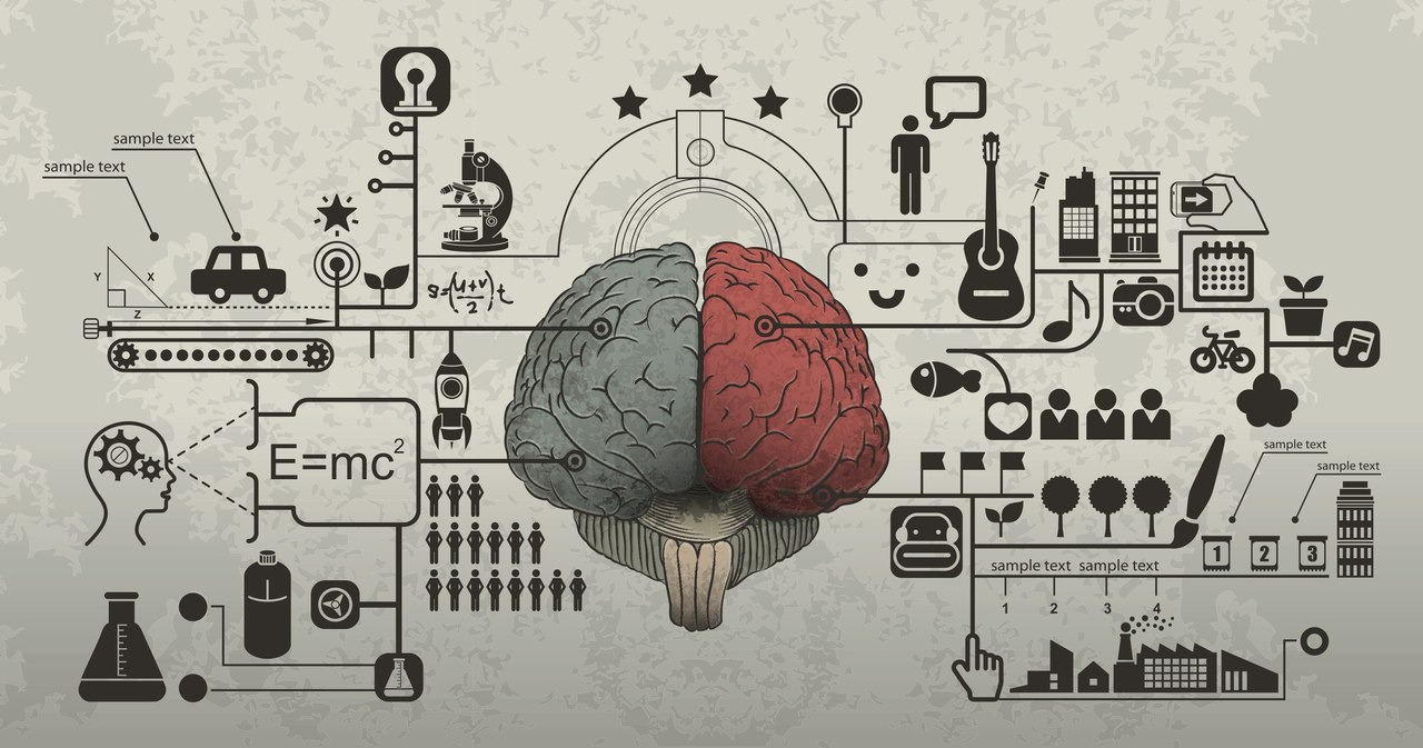
There is such a difference, although it is not limited to the difference between the "creative" right and "logical" left hemispheres. The left hemisphere is largely responsible for speech, verbal thinking, and predicting future events. The right one is for spatial orientation, non-verbal memory and perception of the present. A clear confirmation of the interhemispheric asymmetry of the brain was provided by operations to dissect the cortex of two hemispheres (commissurotomy). But for most mental operations, the coordinated action of both hemispheres is required, and some functions can even be reversed, because the brain is an unusually plastic structure.
It is the ability of the brain to change and rebuild its internal structures. Thanks to this ability, the brain can compensate for damage by "transferring" the performance of certain functions to intact areas: in some cases, a person can lead an almost full life even after the loss of 90% of the neocortex. But the property of neuroplasticity also manifests itself in a more ordinary context - for example, in the learning process. You learn something and even read these lines only because the structure of your brain can change.
Firstly, the volume of gray matter: in men, it is, on average, somewhat larger, although certain zones are more developed in women. Secondly, men tend to have a more developed right hemisphere and parietal cortex, which is probably why they are better at handling spatial operations. In women, the Broca's zone associated with speech is more developed. There are also differences directly related to sexual behavior. But in general, the variations in the structure of the brain between individual women and men are much more significant than the differences between the sexes, so it would be incorrect to talk about a "female" and "male" brain and certain types of intelligence ...
Not true. Each part of the brain performs some function, otherwise it would most likely die off in the process of evolution - why waste energy on a non-working mechanism? Of course, not all parts of the brain work at the same time: they are activated as needed. In addition, lack of mental activity can lead to atrophy of individual cells and increase the susceptibility to Alzheimer's disease. But in general, the brain is an integral structure, in which there are no unnecessary parts. Another thing is that you can use it more or less effectively.
Information from the senses enters the short-term memory, activating neural circuits in the frontal and parietal cortex. Here it lasts only about 30 seconds. For information from short-term memory to pass into long-term memory, repetition is necessary. If a stimulus is repeated long enough, information is consolidated in the hippocampus and supported by neural connections distributed throughout the brain. In some cases, information can be transferred to long-term memory without such mediation, so people with damage to the hippocampus, for example, can improve their driving or drawing skills, although they do not remember anything about training.
The brain is not only a biological, but also a social structure: many features of the psyche are directly related to upbringing and culture. It is believed that representatives of "Western" cultures perceive information, focusing on its individual aspects, and "Eastern" cultures are more sensitive to interconnections and context (hence the connection with the individualism of the former and the collectivism of the latter). There are even correlations between the type of farm (growing rice or wheat) and the type of mental operations. Perception is also influenced by the nature of training, but all these differences are quite specific. There are differences between cultures, but in terms of the psyche, there is still much more in common between them.